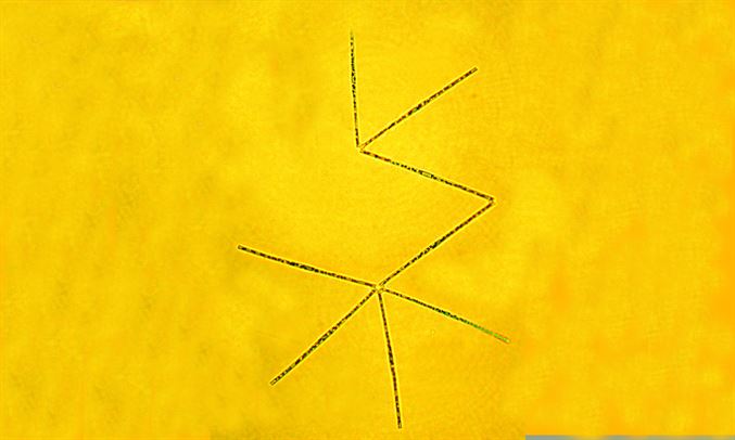
Individual cells measure 80-120 µm in length and 2-4 µm in width. Much like T. nitzschoides, these cells are frequently organized into stellate and/or zigzag colonies. In both valve view and girdle view, the cells generally appear as slender, linear structures. Each end of the cell is rounded, but one of them displays a hollow protrusion, visible in water mounts. These protrusions are exclusive to the valve ends that are not involved in connecting individual cells into colonies. The marginal areolae open into recesses on the valve surface and valve mantle. They are smaller and closely spaced near the valve poles. These areolae have small internal openings and are externally topped by either a simple silica bridge, a triangular arch, or a bridge with marginal flanges. At each valve pole, there is a slit-like labiate process, which can be oriented either obliquely or perpendicularly to the midline of the valve.